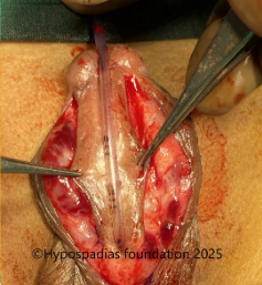
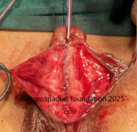
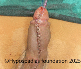
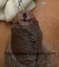
Distal penile hypospadias with severe chordee in an adult – Staged oral mucosa graft repair

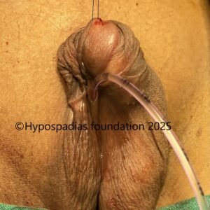
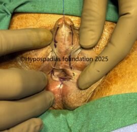
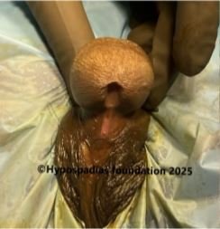
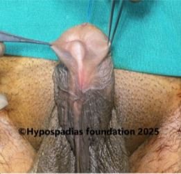
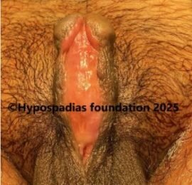
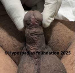
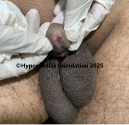
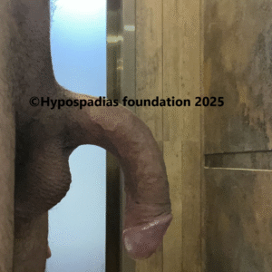
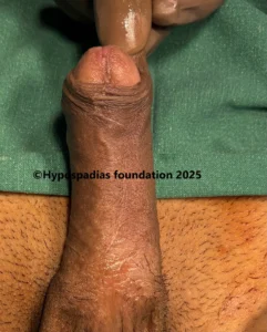
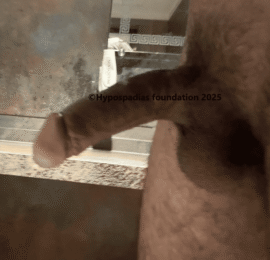
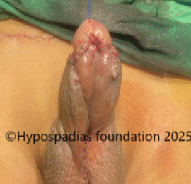
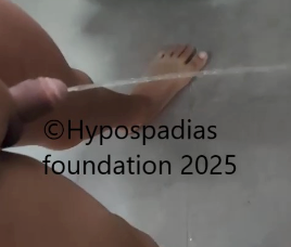
30-year-old male presented to the hypospadias clinic with complaints of passing urine from the distal penile region. He had not undergone any surgery in childhood since his parents were not aware about their child’s hypospadias condition. On examination in the OPD, the meatus was narrow and was in the distal penile region. He also had significant chordee on erection. He was counselled for single or two stage repair depending on the estimation of degree of chordee in the operation theatre.

Stage 1 Repair- Chordee correction and Oral Mucosa graft

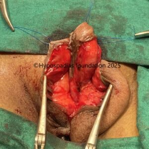
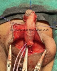
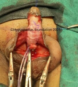
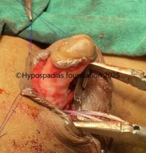
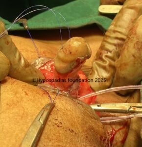
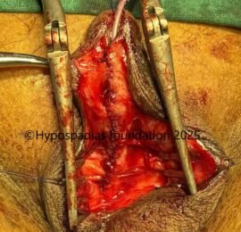
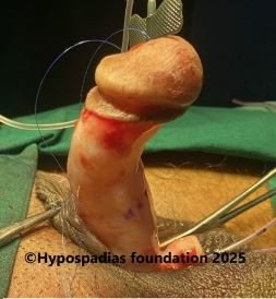
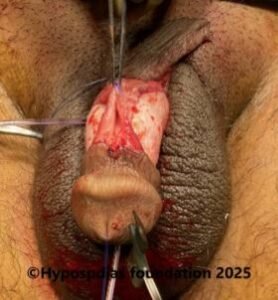
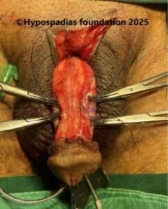
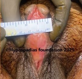
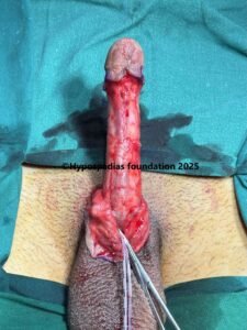
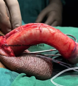
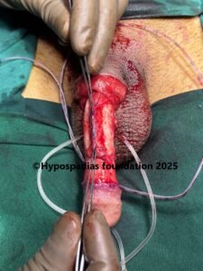
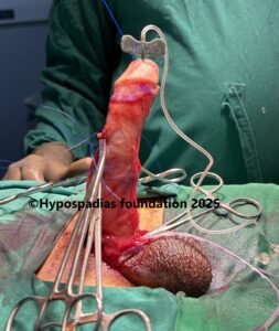
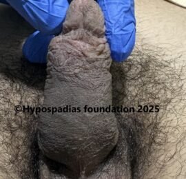
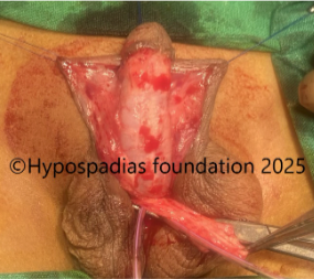
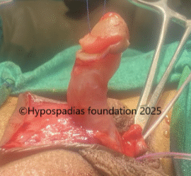
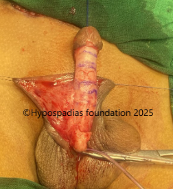
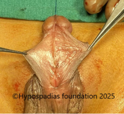
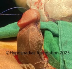
The hypospadias repair surgery was started by complete degloving. Artificial erection test after degloving showed more than 45-degree ventral chordee. Since the chordee was significant with the urethra appearing short and taut, the urethra was divided, and proximal urethra was mobilized. Artificial erection test after urethral plate division showed less than 30-degree chordee, hence we decided to correct it by dorsal 16 dot plication procedure.
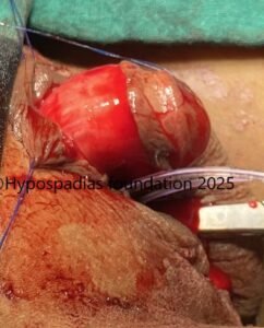
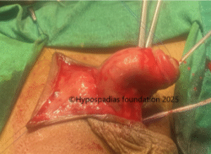
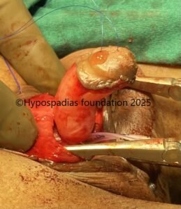
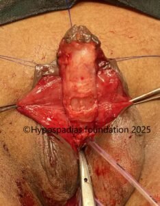
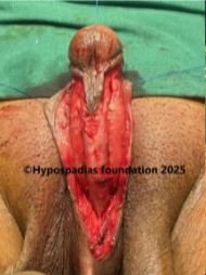
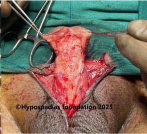
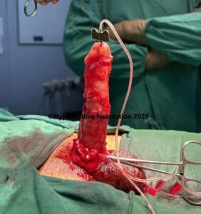
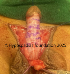
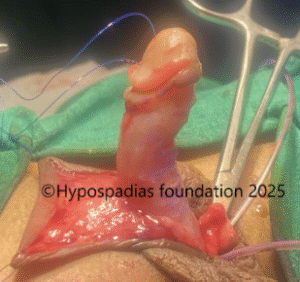
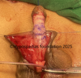
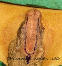
Buck’s fascia along with neurovascular bundle was raised on the dorsal side starting laterally on either side and upto the midline. Artificial erection test was done, and exact site of maximum curvature was marked on the dorsal side. 16 dots (8 dots on either side) were marked- 4 dots proximal and 4 dots distal to the site of maximum curvature. Longitudinal plication sutures were placed at these sites with 4-0 prolene involving full thickness tunica albuginea. Chordee was completely corrected by these equally spaced plication sutures. Buck’s fascia was closed on either side with 6-0 PDS. Proximal urethral stump was fixed at the proximal penile region. Distal tunical bed was prepared for placement of oral mucosa graft. Glans wings were widely raised.

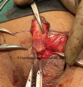
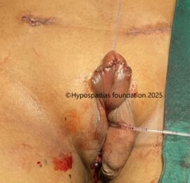
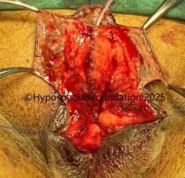
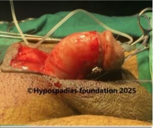
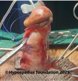
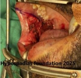
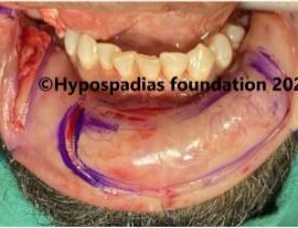
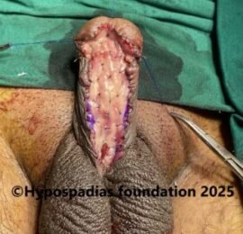
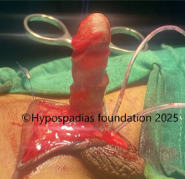
A long 10cm Oral mucosa graft was harvested from inside of the right cheek and lower lip. Graft was defatted and quilted in the urethral bed with 5-0 vicryl. Pressure dressing was done, and the catheter was fixed to the glans stitch. Dressing and catheter were removed after 7 days.

Graft uptake in this case was 100% with no graft loss. After 21 days, local steroid massages were started and continued till 5 months.

Stage 2 Oral Mucosa Graft Urethroplasty

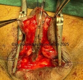
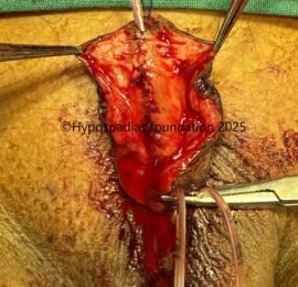
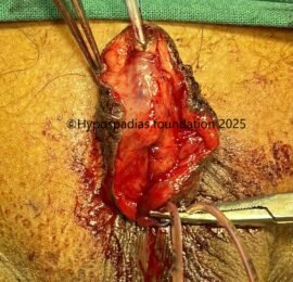
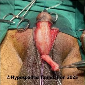
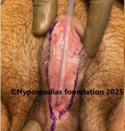
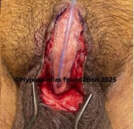
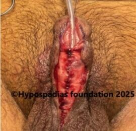
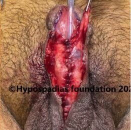
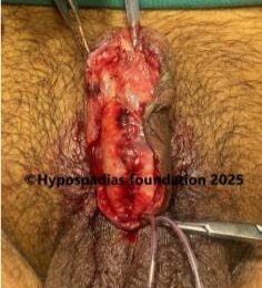
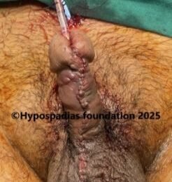
Graft was soft and supple at 6 months after graft placement. Surrounding skin was also heathy and soft. Urethroplasty was started by marking the edges of the graft. Local anaesthesia was infiltrated at the marked site. U-shaped incision was given at the edges of the graft and was deepened till the corpora. Urethroplasty was done over 14Fr silicone catheter with 5-0 vicryl in 2 layers. Local dartos flap was used for second layer coverage. Skin was closed in 2 layers and dressing was done. Suprapubic cystostomy was done with 14Fr malecot’s catheter. Dressing was
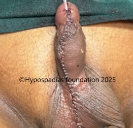
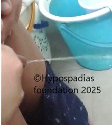
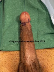
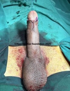
changed on post-surgery day 4 and every 4 th day thereafter. During every dressing change, betadine wash was given, and redressing was done. Per urethral catheter was removed on post operative day 14. Suprapubic catheter was removed on post operative day 21. Patient passed urine in good stream without pain or leak after catheter removal. At 6 months follow up after surgery, the cosmetic result was good, and the patient was passing urine from the glanular meatus in good stream.

43img1
43img2

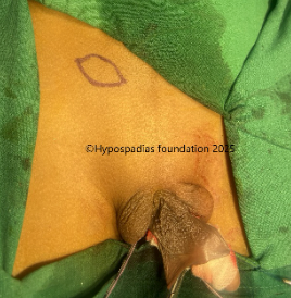
Pic 1: On examination, the meatus was noted to be in the distal penile region

Pic 2: Complete degloving was done. Artificial erection test done showed more than 45 degree chordee

43img6

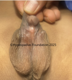
Pic 3: Urethral plate was divided, proximal urethra was mobilized and chordee reassessed. Artificial erection test after urethral plate division showed less than 30 degree chordee.

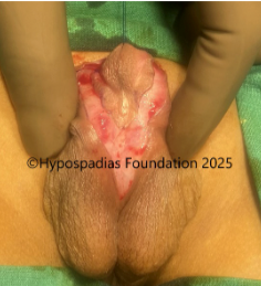
Pic 4: Since the chordee was less than 30 degree, we planned to do 16 dot plication on the dorsal side. Buck’s fascia along with neurovascular bundles were raised and longitudinal plication sutures were placed at 4 points.

Pic 5: 16 dot plication done and chordee was completely corrected. Glans wings were raised

43imf11

Pic 6: Oral mucosa graft harvested from right cheek

43img12

Pic 7: Oral mucosa graft harvested from lower lip

43img13

Pic 8: Oral mucosa graft sutured on the ventral aspect of the penis with 5-0 vicryl thus completing the stage 1 oral mucosa graft placement

43img14
43img15

Pic 9: Assessment at 6 months after stage 1 done. Graft uptake was 100% and graft was soft, supple and ready for stage 2 urethroplasty

43img16
43img17

Pic 10: Marking done at the edges of the graft. Incision given at the edges of the graft and deepened till the corpora.

43img18
43img19

Pic 11: First layer of urtehroplasty done in continuous subcuticular fashion with 5-0 Vicryl. Dratos flap raised from the left side.

43img20
43img21

Pic 12: Dartos flap from the left side sutured over the distal urethroplasty and completion of stage 2 urethroplasty

43img22
43img23

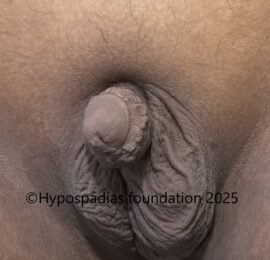
Pic 13: Cosmetic result 6 months after stage 2 showing good outcome and a wide glanular meatus

43img24

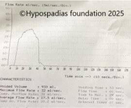
Pic 14: Post surgery uroflowmetry showed normal flow rate

Distal penile hypospadias repair surgery in an adult

Hypospadias is a congenital condition which is present since birth. When not operated in childhood, adults visit our Hypospadias Foundation Clinic for surgical repair in adulthood. The above mentioned case is of a 30 year old male who presented to us with distal penile hypospadias with severe chordee. He was counselled that the decision of single or two stage will be taken during the surgery after assessment of the degree of chordee.

Artificial erection after degloving showed significant chordee and we had to proceed with staged repair. In first stage, the chordee was completely corrected by urethral plate division and dorsal 16 dot plication. We avoid corporotomies in adults because then oral mucosa graft uptake is poor at the sites of deep corporotomy. In adults, the length of oral mucosa graft required is usually long andd hence mostly it needs to be harvested from the cheek and lip. Steroid massages are started on the graft 3 weeks after graft placement.

Second stage is planned 6-8 months after stage 1. Suprapubic cystostomy is a must in all post pubertal and adults undergoing urethroplasty. Post surgery care is extremely important in adults after hypospadias repair. At our centre, we inspect the operated site once every 4 days to make sure that there is no surgical site infection because wound infection after stage 2 can cause dehiscence of the the entire urethroplasty. We remove per urethral catheter first which is around 14 days followed by SPC clamping and removal at 21 days provided there is no dehiscence.

After having done hundreds of adult hypospadias surgeries, we can clearly see differences in adult hypospadias repairs compared to hypospadias repair in a child. Whether it is the size of penis, overall anatomy of tissues, pre-surgery preparation, intra operative sutures or post-op care- everything is very different. Over the years, we have developed various steps which should be done to decrease risk of hypospadias surgery complications in adults. This has led to improved results with more than 95% success rate after hypospadias repair with very few adults requiring another surgery.

At Hypospadias Foundation, a dedicated team of surgeons Dr A.K. Singal and Dr Ashwitha Shenoy take care of adult hypospadias. Dr A.K. Singal is a reputed and top hypospadias surgeon who has been doing hypospadias repair surgeries since 2006 and is rated amongst the best in India and the world for his excellent results. Dr Ashwitha Shenoy is a hypospadias surgeon with experience in managing hypospadias. Both work very closely giving best outcomes to adults with hypospadias. Given their teamwork and dedication to success rates in hypospadias, children and adults come from all over India and more than 20 countries to seek hypospadias treatment under their care.

A closer look at severe chordee correction in an adult by 24 dot plication

25-year-old adult presented to the Hypospadias Foundation OPD with complaints of severe curvature of the penis. There were no urinary complaints. He also that the curvature has worsened over time. On examination, there was severe ventral curvature (downward bending) of the penis. Meatus was located on the glans and was normal in location. No plaques were felt on palpation. Ultrasound doppler of the penis was done which was normal. He was counselled for surgery. Surgery was planned in single or two stage depending on the degree of curvature and the ventral urethral calibre. 

c42img1

Picture 1: Severe curvature of the penis noted on erection

Picture 2: Pre-operative assessment showed normal location of the meatus. Second image shows marking for degloving

Surgery was started by taking a stay stitch on the glans with 4-0 prolene. Marking of the incision was done and local anaesthesia lignocaine with adrenaline was injected at the marked site. Complete degloving was done. Chordee was assessed by artificial erection test and more than 60-degree chordee was noted. Since the penile length was good and urethra was not appearing short or taut, we decided to proceed with dorsal plication procedure to correct the chordee.  

Buck’s fascia along with neurovascular bundle was raised on the dorsal side starting laterally and upto the midline. Chordee was reassessed and exact site of curvature was marked. 24 dots(12 dots on either side) were marked. 4 dots proximal and 8 dots distal to the site of maximum curvature. Longitudinal plication sutures were placed at these sites with 4-0 prolene involving full thickness tunica albuginea. Chordee was reassessed and there was no chordee. Chordee was completely corrected by these plication sutures. Buck’s fascia was closed on either side with 6-0 PDS. Circumcision was completed and skin rearranged and closed in 2 layers with 6-0 PDS and 5-0 vicryl rapide. 12Fr silicone catheter was placed per urethra and dressing was done. Catheter was removed after 7 days. Follow up at 6 months after surgery, there was no chordee and the erection was straight. 

case42img4

Picture 3: Complete degloving done and artificial erection test done which showed more than 60-degree chordee.

Picture 4: Neurovascular bundle raised on both sides upto the midline. Marking done, plications sutures placed, held and artificial erection test done. No chordee noted

Picture 5: 24 dot plication completed and circumcision done

c42img10 (1)

Picture 6: Penile  erection straight 6 months after surgery

c42img11

Picture 7: Cosmetic result 6 months after surgery

Severe ventral chordee in adults: Diagnosis and management

Chordee is characterized by curvature of the penis which is noticeable only during penile erection. This is congenital (present at birth) but can be missed in childhood and can present with challenges in adulthood potentially causing discomfort, affecting sexual function and can also have psychological implications impacting self-esteem.

In chordee, there is noticeable bend in the penis during an erection which can be downward, upward or sideways. It can cause pain or discomfort during erection or sexual intercourse. Surgical correction is the only effective method of chordee correction in adults. The goal of the surgery is to straighten the penis and restore normal function. Chordee correction can be done by dorsal plication or ventral lengthening procedure. Ventral lengthening procedure is done if there is short urethra which contributes to the penile curvature. If the urethra is normal with good penile length, then dorsal plications are preferred. 

In the above-mentioned case, the penile length was good, and the urethra was normal hence we proceeded with dorsal plication. In this procedure multiple (16 or 24) non absorbable fine sutures are strategically placed on the convex side of the penis. These sutures are placed in pairs and in parallel fashion to create plications in the tunica albuginea. Neurovascular bundles are carefully raised before placing the plication sutures. 

Important note for adult chordee

It is essential for adults considering chordee correction to have a thorough evaluation by a hypospadias specialist who specialized in penile reconstructive surgery. The surgeon will assess the type and degree of curvature, discuss the case and recommend the most appropriate surgical technique whether plication or a staged procedure. 

At hypospadias foundation, we get adults and children from all over the world in search of treatment for hypospadias. We provide support and information for children, adults and their families affected by hypospadias. Dr Singal and Dr Shenoy are deeply devoted to creating awareness and helping patients get the right treatment for hypospadias be it primary, redo or adult hypospadias. Children and adults from more than 25 countries visit our hypospadias foundation in search for cure and are cured of hypospadias. 

Dr A K Singal is a highly experienced surgeon and regarded as the best hypospadias surgeon in India and in the world. He has dedicated his life towards treating children and adults with hypospadias. His expertise in this area has helped us achieve excellent outcomes in adults and children with hypospadias.

Dr Ashwitha Shenoy is an expert pediatric surgeon with special interest in pediatric urology and hypospadias. Both Dr Singal and Dr Shenoy work together to give

Two stage repair of deceptive distal penile hypospadias with hypoplastic urethra and severe chordee

6-year male from Kerala presented to the Hypospadias Foundation clinic with complaints of passing urine from distal penile region associated with downward curvature of the penis (chordee). On examination, meatus was in distal penile region, but the urethra was thin till the proximal penile region. The stretched penile length (SPL) was 40mm and glans diameter (GD) was 12.5mm. In view of small glans diameter, he received two doses of injection testosterone in the pre surgery period which were given 1 month apart. Following the two doses, the stretched penile length improved to 35mm and glans diameter to 15mm.

He was planned for hypospadias repair and the family was counselled for single or two stage repair based on the degree of chordee.

The surgery was started by complete degloving which means the penile skin was completely taken down preserving its blood supply. After degloving, the chordee was assessed by artificial erection test. Here in this case after complete degloving, artificial erection test showed chordee of more than 45 degrees. This was because of the hypoplastic (underdeveloped) tissues on the underside of the penis. We decided to divide the urethral plate and mobilise the urethra. Spongiosum was raised on the sides first, urethral plate divided at coronal level and urethra mobilised till penoscrotal junction to fully evaluate the chordee. Chordee was reassessed and there was persistent chordee of more than 45 degrees. Site of maximum curvature was marked on the ventral side (underside of the penis) and three corporotomies were made by placing horizontal incisions over the tunica with middle incision at the site of maximum curvature. Chordee was reassessed and there was still significant chordee necessitating a ventral penile lengthening procedure called as dermal graft. The central corporotomy incision was deepened to full divide tunica and then cavernosal tissue was mobilised from tunica on all sides. A skin ellipse was harvested from the inguinal region, de-epithelized, and defatted to create a dermal graft. This dermal graft was sutured at the site of tunical defect with 6-0 PDS interrupted sutures. An artificial erection test following dermal graft revealed no chordee.

Once the chordee was corrected, the proximal urethra was sutured in the proximal penile region with 6-0 PDS. Glans wings were raised. Byars flap was created. Byars flap involves division of the dorsal prepuce in the midline and then rotation to the underside of the penis to cover the whole underside of the penis from glans till all around the meatus.

41img1
41img2

Picture 1: On clinical examination, the meatus was in the distal penile region with thin urethra till the proximal penile region. Surgery was started by complete degloving

Picture 2: Artificial erection test showed chordee of more than 45 degrees

41img5

Picture 3: Urethral plate divided, mobilised and chordee reassessed. More than 45 degrees chordee noted

Picture 4: Three ventral corporotomies given and chordee reassessed, persistent chordee of more than 30 degrees noted, hence planned to go ahead with ventral lengthening procedure

Picture 5: Deep corporotomy given at the site of maximum curvature and tunica incised, dermal graft harvested from the right inguinal region

41img10
41img11

Picture 6: Dermal graft sutured at the site of tunical defect. No chordee noted after dermal graft.

41img12

Picture 7: Dorsal prepuce (foreskin) divided in the midline and flaps rotated to cover the underside of the penis

Second stage of Hypospadias Repair:

After 6 months of first hypospadias surgery, the child was taken up for second stage urethroplasty. We checked chordee again before starting the urethroplasty (urethral reconstruction) and the penis was completely straight. The prepucial flaps which were done in first stage had healed very well. The flaps were wide and supple. 15mm wide incision was marked on the flaps and, local anaesthesia was infiltrated at the marked site and incisions were deepened till the corpora. Urethroplasty was done over 7Fr Infant feeding tube in two layers with 6-0 PDS absorbable sutures. Dartos flap was harvested from both sides and sutured over the entire urethroplasty. Glans wings were raised. Distal urethroplasty and glansplasty was done, and a new meatus was created on the glans. The dressing in the post operative period was changed on post operative day 7 and catheter was removed on post operative day 12. Child was passing urine through the new opening in a good stream.

41img13
41img14

Picture 8: Prepucial flaps 6 months after stage 1 were healed very well. Flaps were wide and supple. Artificial erection test done before urethroplasty done showed no residual chordee

41img15
41img16

Picture 9: 15mm wide flaps marked and incised.

41img17

Picture 10: Urethroplasty done over 7Fr Infant feeding tube, dartos flap raised and sutured over the urethroplasty and glansplasty done

41img18
41img19

Picture 11: Completion of urethroplasty and follow-up at post operative day 12

41img20

Picture 12: Urine stream following catheter removal

Distal penile hypospadias with severe chordee (Deceptive hypospadias)

In some cases of hypospadias, the urethral opening is not only in an abnormal location but also the urethra can be underdeveloped or hypoplastic. Hypoplastic urethra is characterized by thin, underdeveloped, paperlike quality of urethral plate and the existing distal urethra. While the meatal opening may be near the tip, the presence of a hypoplastic or underdeveloped urethra indicates a more complex form of hypospadias, even if the urethral meatus appears relatively distal. This is often associated with chordee which contributes to the overall complexity of the hypospadias.

The hypospadias repair in hypoplastic urethra is more challenging. This thin urethra does not have enough robust tissue to create a new functional urethra. Using this thin urethra in reconstruction can lead to complications such as fistula formation, stricture, diverticulum formation or dehiscence.

The surgical management in hypoplastic urethra often requires specialized techniques. Degloving in these cases must be done carefully because of higher chance of injury to the urethra due to its paper-thin consistency. The type of surgical repair in cases with hypoplastic urethra is decided based on the degree of chordee. If the chordee is not significant and if single stage repair is planned, then either the thin urethra must be laid open before staring the urethral reconstruction or spongiosa around the thin urethra has to be mobilized and sutured over the thin urethra which is called as Spongioplasty.

For severe cases, like in the above-mentioned case with significant chordee and extensive hypoplastic urethra, two stage repair may be necessary. In the first stage, the penis is straightened and prepucial flaps are created. In the second stage which is 6 months after stage 1, the flaps are tubularized to form the new urethra.

Long term follow in cases of hypoplastic urethra is necessary because complications like stricture can develop years after hypospadias repair surgery. Hence these complex cases of hypoplastic urethra with hypospadias should be performed by an experienced hypospadias surgeon.

At hypospadias foundation, we have operated many cases of hypospadias with hypoplastic urethra. Each case is thoroughly examined in the preoperative period, carefully degloved during the surgery, meticulously assessed and hypospadias repair is done considering factors such as degree of chordee, severity of hypoplasia and quality of urethral plate. This has helped us achieve success rate of more than 95% even in the most complex cases.

The team at Hypospadias Foundation has two experienced hypospadias surgeons. Dr A.K Singal is considered the best hypospadias surgeon in India and the world and has a special expertise in managing complex hypospadias cases. His profound anatomical knowledge, meticulous surgical skill and an unwavering commitment to restoring both form and function in hypospadias has transformed various lives.

Dr Ashwitha Shenoy is an expert hypospadias surgeon who along with Dr Singal has dedicated her life to treating children and adults with hypospadias. Their dedication extends beyond the operating room, offering compassionate guidance and hope to families navigating a challenging diagnosis.

Staged repair of Midpenile hypospadias with severe chordee

2 year 6 months male child came to Hypospadias Foundation from Nepal with complaints of passing urine from the mid penile region associated with abnormal ventral curvature of the penis. Parents were looking for an experienced hypospadias surgeon hence they delayed the surgery till 2.5 years of age. On examination, the meatus was in the mid penile region with poor urethral plate. He was planned for hypospadias repair, and we counselled the family that whether the surgery would be in 1 stage or two stages will be decided depending on the degree of chordee during surgery.

At the start of hypospadias surgery, the entire penile skin was taken down all around till the base of the scrotum, a step known as complete degloving. Chordee was assessed after complete degloving. Chordee was noted to be more than 45 degrees. This was because of deficient tissues on the underside of the penis. We made a horizontal incision on the underside of the penis at the site of maximum curvature and divided the urethral plate. After division of the urethral plate, the proximal urethra was mobilized. Chordee was reassessed and there was persistent chordee of more than 30 degrees noted. Site of maximum curvature was marked on the ventral side. Three Corporotomies were done by placing horizontal incisions ventrally over the tunica with middle incision at the site of maximum curvature. Chordee was reassessed and less than 30-degree chordee was noted. Complete Chordee correction was achieved by tunica albuginea plication at 12’o clock position on the dorsal side. Artificial erection test showed straight penis.

After that the foreskin(prepuce) was divided in the midline and rotated to cover the underside of the penis- a step known as Byar’s flaps. This hairless prepucial skin which is now on the underside of penis will be utilized in the second stage for urethroplasty.

40img1
40img2

Picture 1: On clinical examination, the meatus was at the mid penile region with severe chordee

40img4

Picture 2: Marking done for degloving and complete degloving done

40img6

Picture 3: Artificial erection test was done which showed chordee of more than 45 degrees and hence urethral plate division was done

Picture 4: Persistent chordee of more than 30 degrees hence three ventral corporotomies given

Picture 5: Less than 30 degrees chordee noted after three ventral corporotomies hence 12’o clock dorsal tunica albuginea plication done. No chordee after dorsal tunical plication

40img11
40img12

Picture 6: Dorsal prepuce(foreskin) divided in the midline and rotated to cover the underside of the penis

Second stage of Hypospadias Repair:

After 6 months of first hypospadias surgery, the child was taken up for second stage urethroplasty. We checked chordee again- the penis was completely straight. The prepucial flaps which were done in first stage had healed very well. The flaps were wide and supple. 15mm wide incision was marked on the flaps and, local anaesthesia was infiltrated at the marked site and incisions were deepened till corpora and hemostasis checked. Urethroplasty was done over 7Fr Infant feeding tube in two layers with 6-0 PDS absorbable sutures. Glans wings were raised, Glansplasty was done, and a new meatus was made on the glans. The dressing in the post operative period was changed on post operative day 7 and catheter was removed on post operative day 10. Child was passing urine through the new opening in good stream.

40img13
40img14

Picture 7: Prepucial flaps 6 months after stage 1 were healed very well. Flaps were wide and supple.

40img15

Picture 8: Artificial erection test done – completely straight penis noted

40img16
40img17

Picture 9: Urethroplasty done over 7Fr Infant feeding tube and dartos flap raised on both sides

40img18
40img19

Picture 10: Glans wings raised, distal urethroplasty done and Dartos flap sutured over the urethroplasty.

40img20

Pic 11: Glansplasty and skin closure done, completing Stage 2 urethroplasty

40img21

Pic 12: Post catheter removal, passing urine in single straight stream with no complications.

Two stage repair in hypospadias with severe chordee

Staged repair in hypospadias is a preferred approach in cases of severe hypospadias when the urethral opening is significantly displaced and there is significant chordee. Two stage repair involves two separate surgeries to correct the hypospadias, the first stage being correction of the bend of the penis and rearranging the foreskin on the underside of the penis. The second stage is usually performed 6-12 months after stage 1 hypospadias repair.

Two stage repairs are often considered to offer superior functional and cosmetic results in severe hypospadias compared to attempting a single stage complex repair. They allow for a
more controlled approach to address significant curvature and create a neourethra with potentially better tissue quality from the prepucial flaps. Whether your child needs a single stage repair or a staged approach is best decided by the expert hypospadias surgeon during the initial steps of surgery and is primarily dictated by the degree of curvature.

About Hypospadias foundation

Hypospadias foundation is an organization dedicated for children and adults with hypospadias and is rated as the best hospital for hypospadias repair in India and the World. Hypospadias foundation offers best results for surgical repair for primary and failed(redo) hypospadias. We at hypospadias foundation believe in providing personalized care and psychosocial support to families of children and adults with hypospadias. Dr A K Singal is a well renowned hypospadias surgeon and regarded as the best hypospadias surgeon in India and in the world. His vision for hypospadias has helped him achieve excellent outcomes in adults and children with hypospadias. He has dedicated his life in treating children and adults with hypospadias with his innovative surgical techniques. Dr Ashwitha Shenoy is an expert pediatric surgeon with special interest in pediatric urology and hypospadias. She holds a particular interest in hypospadias and along with Dr Singal performs advanced surgical techniques for both primary and complex hypospadias cases in children and adults.

Contact Us

Call Now