
A 25-year-old man from Assam, India presented to the Hypospadias Foundation with complaints of passing urine through an opening located in scrotal region. Clinica examination revealed severe scrotal hypospadias associated with significant ventral penile curvature (chordee) and penoscrotal transposition. The patient reported longstanding psychological concerns regarding urinary function, penile appearance, and the impact of the condition on his confidence and future sexual function.
After detailed clinical evaluation and counselling, a staged reconstruction of scrotalhypospadias was planned. Because of the severity of the deformity and the degree of curvature anticipated, the surgical team proposed a three-stage approach consisting of chordee correction, urethral reconstruction, and later correction of penoscrotal transposition. The patient was counselled regarding the planned procedures, expected outcomes, and potential complications.
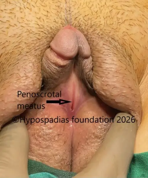
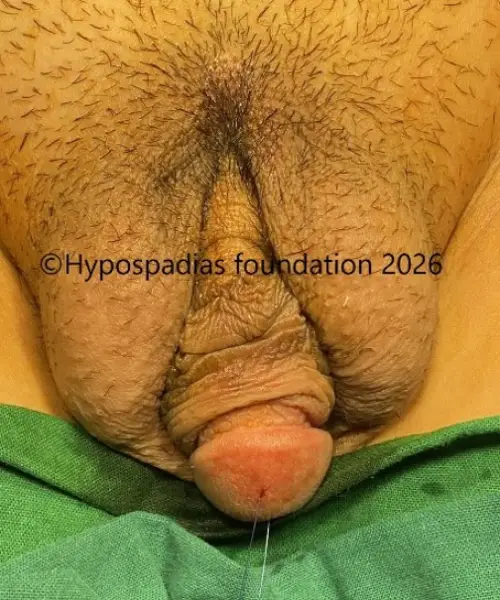
Pic 1 & 2: Clinical examination showing penoscrotal meatus and penoscrotal transposition
Stage 1 chordee correction:
Under general anesthesia, the patient was prepared and draped in the standard sterile fashion. Marking of the degloving incision was performed. Complete penile degloving was carried out circumferentially up to the level of the suspensory ligament dorsally and the scrotal fat pad ventrally.
An artificial erection test was performed by inserting a butterfly needle into the glans penis and injecting sterile saline into the corpora cavernosa. Severe ventral curvature exceeding 45° was noted. Mobilization of the corpus spongiosum was performed on both sides. The urethral plate was divided, and the proximal urethra was mobilized. Repeat artificial erection testing demonstrated persistent ventral curvature of more than 45° despite urethral plate division.
Surgical Decision-Making
At this stage, the persistence of severe curvature despite complete urethral plate division confirmed that the chordee was not solely caused by tethering of the urethral plate. In our experience, residual curvature of this magnitude requires additional straightening procedures to achieve a functionally straight erection and prevent future sexual difficulties. A dorsal 24-dot plication was therefore selected to provide reliable correction while preserving erectile function.
In view of the persistent severe chordee, dorsal 24-dot plication was planned. The point of maximum curvature was identified and marked. Equidistant sites for plication sutures were marked on either side of the dorsal penile shaft. Buck’s fascia along with the neurovascular bundle was carefully elevated from lateral to medial to expose the tunica albuginea at the site of correction.
Three plication sutures were placed on either side of the midline (total 24-dot plication). The sutures were temporarily secured with rubber shods, and a repeat artificial erection test was performed. Complete correction of the chordee was achieved. The plication sutures were then tied securely.
The proximal urethral stump was fixed at the penoscrotal junction. The dorsal preputial skin was divided in the midline and transposed ventrally as Byars’ flaps for ventral skin coverage. A 14 Fr silicone urethral catheter was inserted and secured.
Postoperatively, dressing changes were performed on postoperative days 4 and 7. The urethral catheter was removed on postoperative day 10. The patient tolerated the procedure well and had an uneventful postoperative recovery.
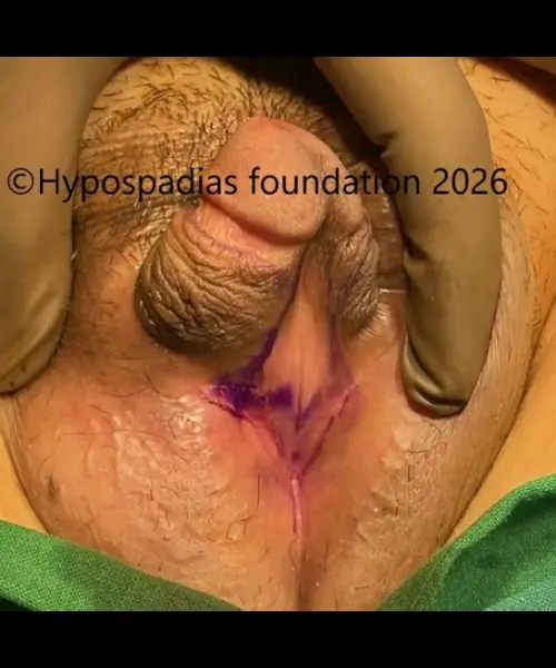
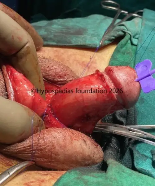
Pic 3 & 4: Marking of the degloving incision and chordee assessment by artificial erection test which showed more than 45 degree chordee
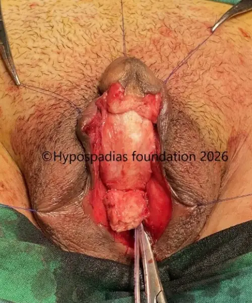
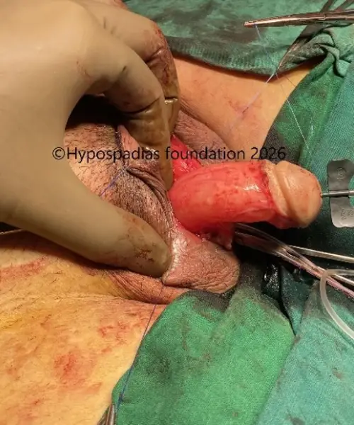
Pic 5 & 6: Urethral plate division, proximal urethral mobilization. Chordee assesment after urethral plate division showed persistent chordee of more than 45 degree
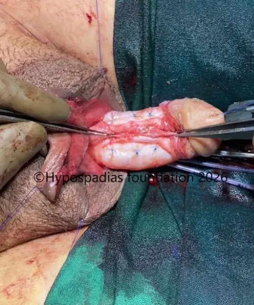
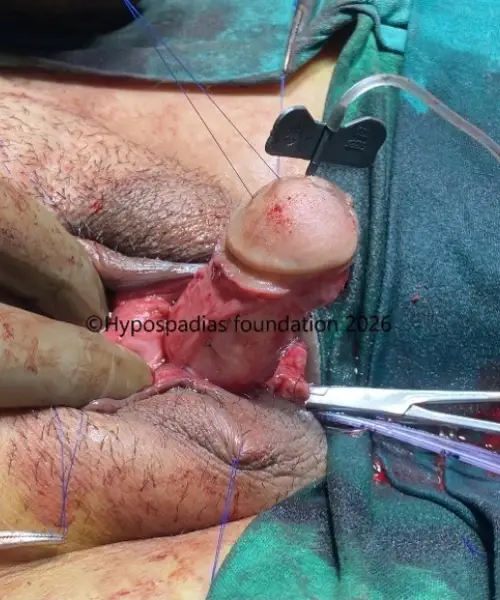
Pic 7 & 8: 24 dot tunical albuginea plication done following which there was no chordee
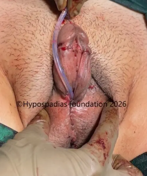
Pic 9: Dorsal prepuce divided in the midline and skin rearranged as byars flap. 12Fr silicone catheter inserted per urethra.
Second stage urethroplasty and outcome:
At 6-month follow-up after the first-stage surgery, the Byars' flaps were healthy and well vascularized with excellent healing. Although the patient had initially been planned for a three-stage reconstruction because of the severity of the deformity, the excellent condition of the tissues allowed progression to urethroplasty during the second stage itself.
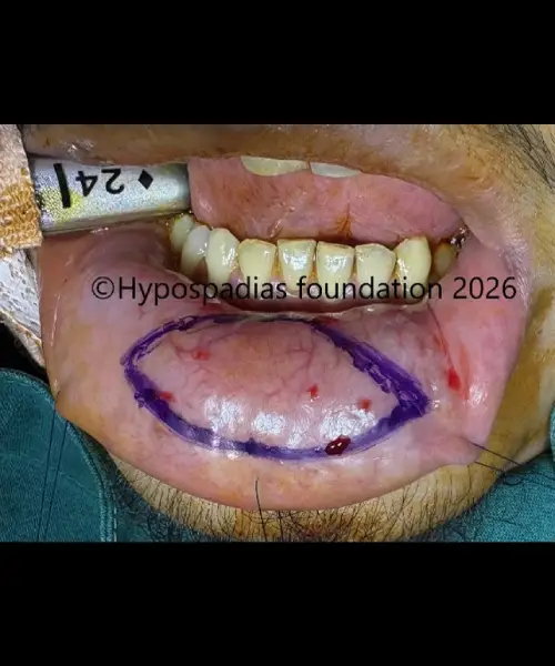
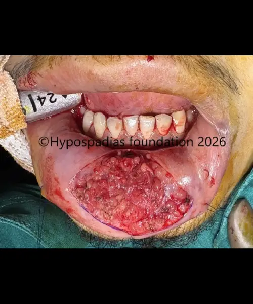
During the second-stage procedure, the proximal segment of the neourethra from the penoscrotal junction to the mid-penile shaft was reconstructed using the Byars’ flap that had been transposed during the first-stage surgery. For the distal segment extending from the mid penile region to the glans, an oral mucosal graft was used as an inlay graft. The graft, measuring 25 × 10 mm, was harvested from the lower lip.
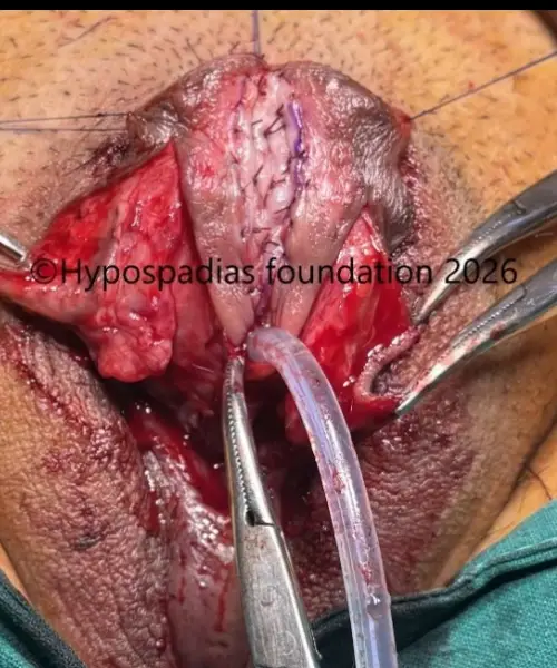
The oral mucosal graft was quilted to the underlying corpora cavernosa in the midline and secured to the surrounding tissue edges. A urethral plate width of 20 mm was marked, and the edges were incised and deepened to the corporal bodies. Tubularized urethroplasty was then performed over a 12 Fr silicone catheter in two layers: the first layer using continuous subcuticular sutures and the second reinforcing layer using interrupted 5-0 Vicryl sutures.
A well-vascularized dartos flap was raised from both sides and placed as an additional protective layer over the urethroplasty. Skin closure was completed in two layers using 5-0 Vicryl and 5-0 Vicryl Rapide sutures. A suprapubic cystostomy was performed at the end of the procedure for urinary diversion.
Postoperatively, dressings were changed every fourth day. The per-urethral catheter was removed on postoperative day 18. The suprapubic catheter was clamped on postoperative day 20, and the patient demonstrated a good urinary stream. The suprapubic catheter was subsequently removed.
Functional Outcome
The patient experienced an uncomplicated postoperative recovery. Following catheter removal, he demonstrated a good urinary stream without evidence of fistula formation, wound breakdown, or urethral stricture. Clinical assessment confirmed successful healing of the reconstructed urethra and maintenance of penile straightness.
Although a three-stage reconstruction had originally been anticipated because of the severity of the deformity, the excellent healing and vascularity of the first-stage repair allowed completion of reconstruction during the second stage. This reduced the overall treatment burden while achieving an excellent functional outcome.
The patient expressed satisfaction with the urinary function and appearance achieved following reconstruction.
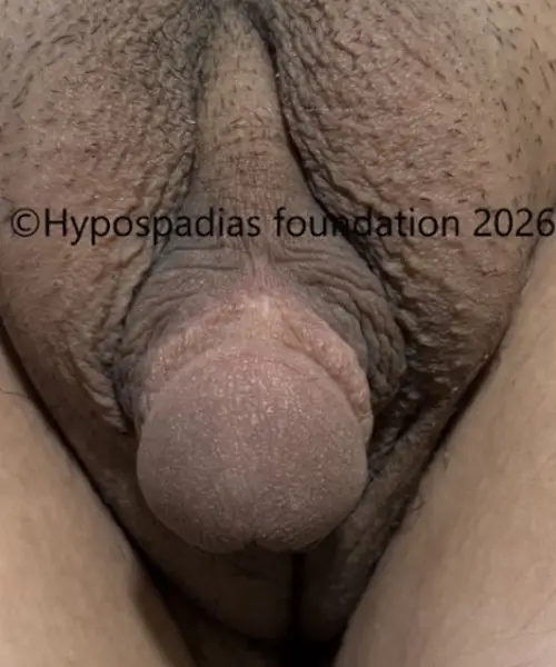
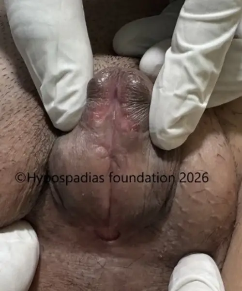
Pic 10 & 11: 6 months after stage 1, no chordee noted and the flaps were healthy
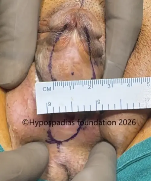
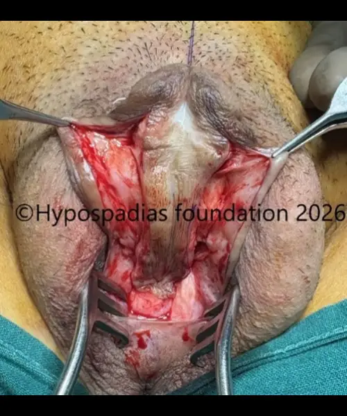
Pic 12 & 13: 20mm wide marking of the flaps done, incision given at the marked site and deepened till the corpora
Pic 14 & 15: Proximal urethroplasty done upto the midpenile region from the byars flap placed in the first surgery. Midline incision given distally for placement of oral mucosa as an inlay graft
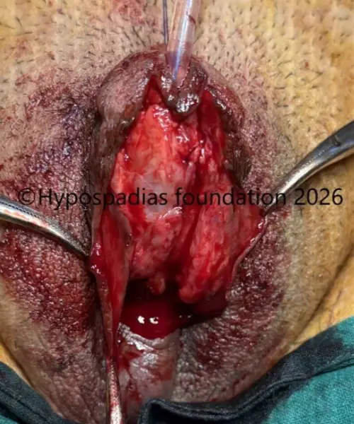
Pic 15 & 16: Oral mucosa graft harvested from the lower lip of size 25x10mm
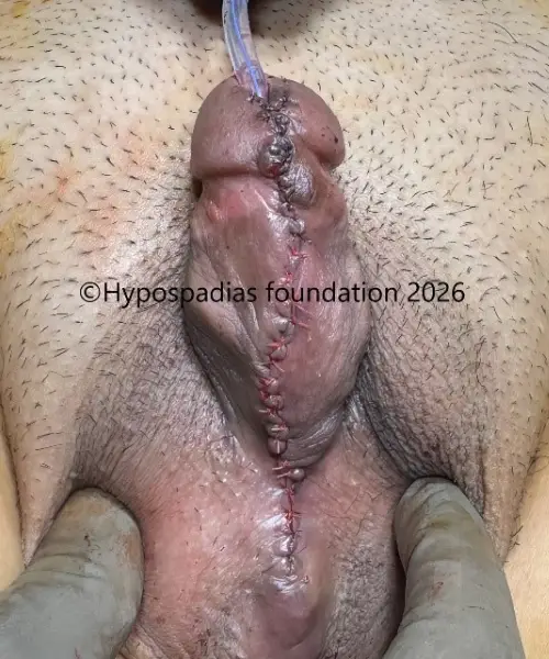
Pic 17: Glansplasty and skin closure done. Suprapubic cystostomy done at the end of the surgery
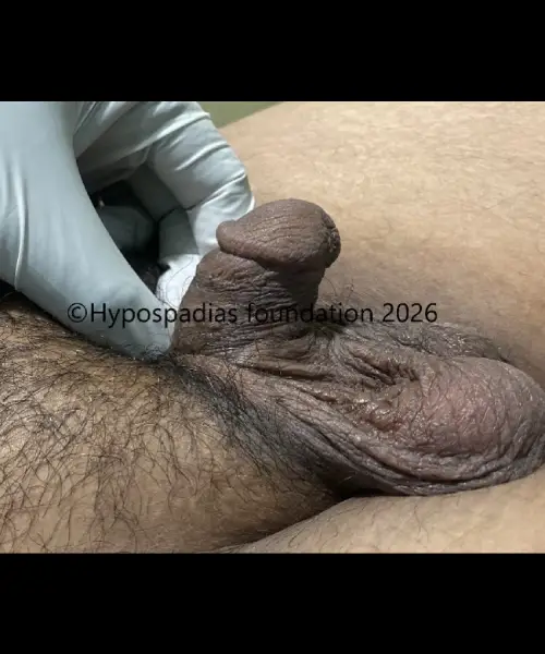
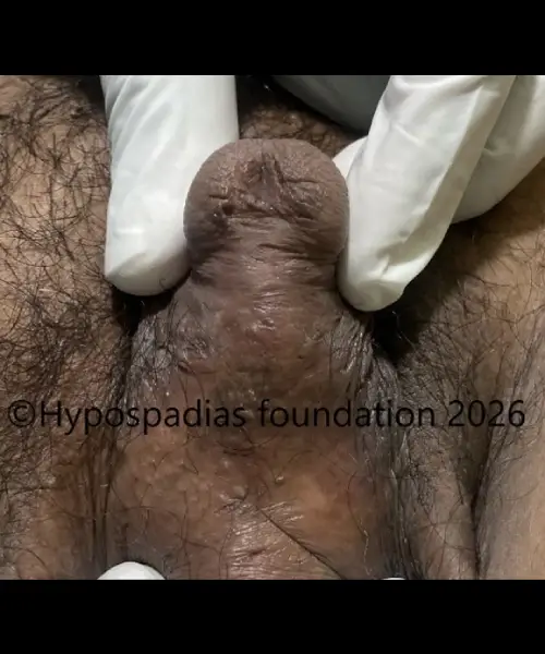
Pic 18 & 19: Cosmetic result at 6 months after stage 2 shows straight penis with meatus on the glans
Severe Hypospadias in an Adult Successfully Treated with a Two- Stage Repair
Hypospadias is a congenital condition in which the urinary opening (meatus) is located on the underside of the penis rather than at its tip. In severe forms of hypospadias, the opening may be located at the penoscrotal junction, within the scrotum, or even in the perineal region. These cases are often associated with significant penile curvature (chordee).
Many patients with severe hypospadias undergo surgery during childhood. However, some individuals reach adulthood without treatment and seek correction later in life because of functional, sexual, and psychological concerns.
Common Symptoms in Adults with Severe Hypospadias
Adults with untreated severe hypospadias commonly present with:
Having to sit down to pass urine as urine goes backwards
Spraying of the urinary stream
Severe penile curvature during erection
Painful erections or difficulty during sexual intercourse
Concerns regarding penile appearance
Reduced self-confidence and psychological distress
Challenges of Adult Hypospadias Repair
Adult hypospadias surgery presents unique challenges that are less commonly encountered in children. Over many years, untreated penile curvature may become more pronounced, making correction technically more demanding. Adult tissues are generally less elastic, wound healing may be slower, and postoperative erections can place additional stress on the surgical repair.
In our practice, adults with severe penoscrotal hypospadias frequently require staged reconstruction to safely achieve complete straightening of the penis and creation of a durable urethra. Complete correction of penile curvature is essential to restore both function and sexual confidence. Careful surgical planning and meticulous tissue handling are critical to achieving successful outcomes while minimizing complications. In many adult patients, oral mucosal grafts are required because local tissues alone may not be sufficient for reconstruction.
The present case demonstrates that withindividualized reconstruction strategies we can achieve good outcomes even in severe adult hypospadias to be corrected successfully.
Psychological Impact of Untreated Adult Hypospadias
The effects of severe hypospadias often extend beyond urinary and sexual function. Many adults report years of embarrassment related to penile appearance, anxiety regarding intimate relationships, and reduced self-confidence. Some avoid seeking treatment because they are unaware that effective surgical options remain available in adulthood.
Addressing these concerns is an important component of treatment. Counselling, realistic expectation setting, and comprehensive follow-up can help patients regain confidence alongside physical recovery.
Key Learning Points from This Case
Severe adult hypospadias can be successfully corrected with staged reconstruction.
Complete correction of penile curvature is essential for long-term functional outcomes.
Oral mucosal grafts remain an important option when local tissues are insufficient for urethral reconstruction.
Careful surgical planning can sometimes reduce the number of planned stages without compromising outcomes.
Adults with untreated hypospadias can achieve excellent urinary, cosmetic, and psychological outcomes with expert surgical care.
Adult Hypospadias Treatment at Hypospadias Foundation
At Hypospadias Foundation, we regularly evaluate and treat adults with both primary and failed hypospadias repairs. Our team specializes in advanced reconstructive techniques, including single-stage and staged repairs using oral mucosal grafts when required. We understand the physical and emotional challenges associated with adult hypospadias and provide comprehensive care throughout the treatment
journey.
With careful evaluation, individualized treatment planning, and expert surgical care, adult men with hypospadias can achieve excellentfunctional and cosmetic outcomes and regain confidence in their daily lives.
Expert Hypospadias Surgeons in India
Dr. A. K. Singal is a highly experienced hypospadias surgeon who has dedicated his career to the management of primary, complex, and failed hypospadias repairs in children and adults. His clinical practice focuses on advanced reconstructive techniques for severe hypospadias and urethral reconstruction.
Dr. Ashwitha Shenoy is a pediatric surgeon and urologist with a special interest in pediatric urology and hypospadias surgery. She works closely with Dr. Singal in the management of both primary and complex reconstructive cases.
Together, they provide comprehensive care for patients with hypospadias, including detailed evaluation, individualized treatment planning, advanced reconstructive surgery, and long-term follow-up.